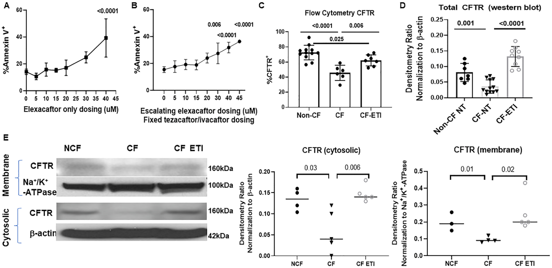
Figure 1: ETI treatment is associated with increased MDM CFTR expression.

A) Flow cytometry measurements of cellular apoptosis by detection of % Annexin V positive CF MDMs in response to escalating doses of elexacaftor (0–40 μM). 40μM elexacaftor significantly increased MDM apoptosis, p <0.0001 via one-way ANOVA with Tukey’s test, n= 4–6. B) Flow cytometry % Annexin V positive CF MDMs in response to escalating doses of elexacaftor combined with fixed tezacaftor and ivacaftor dosing (5μM). Elexacaftor doses above 30μM were associated with significantly increased MDM apoptosis, p = 0.006 and <0.0001 via one-way ANOVA with Tukey’s test, n=4–7. C) Quantitation of CFTR expression via flow cytometry detection of %CFTR+ fluorescence in non-CF (n=12), CF (n=6), and CF-ETI (n=7) MDMs, detected via UNC-596 or Alamone ACL-006 antibody. P values for individual comparisons are shown, one-way ANOVA. Negative and single-color controls not shown. D) Densitometric ratios of western blot analysis of CFTR (UNC-596 antibody) Band C expression in non-CF (n=7), CF (n = 12), and CF-ETI (n=9) MDMs. Densitometry ratios normalized to the loading control β-actin. P values for individual comparisons are shown, one-way ANOVA, non-CF vs CF-ETI non-significant. E) Subcellular fractionation western blot of membrane and cytosolic fractions of CFTR in non-CF, CF, and CF-ETI MDMs. Na+/K+-ATPase was used as a membrane control, and β-actin as a cytosolic control. Representative image is shown with corresponding densitometry for cytosolic and membrane fractions from all replicates, unpaired t-tests.
