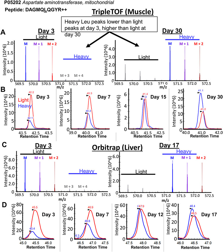
Figure 3.
Heavy leu incorporation is observable in Skyline software. (A) The isotope envelope of the representative peptide DAGMQLQGYR from aspartate aminotransferase, in mouse skeletal muscle detected on a triple quadrupole time-of-flight instrument after 3 (left) or 30 days (right) on a synthetic diet. Incorporation of heavy leucine is clearly visible in Skyline by the increase in abundance of the “heavy” leucine isotopic peaks. (B) Extracted ion chromatograms of the peptide DAGMQLQGYR over four time points shows an increase in the peak area of deuterated heavy leucine (blue, 571.2785 m/z), relative to light leucine (red, 569.7691 m/z), over time. The equivalent (C) isotopic envelope and (D) extracted ion chromatograms for DAGMQLQGYR in liver lysates from an Orbitrap are shown.

