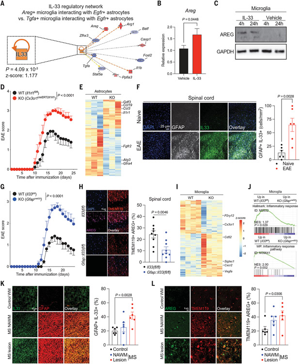
Fig. 4. IL-33-ST2 signaling controls an astrocyte–microglia regulatory circuit.
(A) IL-33 regulates Areg+ microglial interactions with Egfr+ astrocytes determined by RABID-seq during peak EAE. (B and C) IL-33 induces the expression of Areg/AREG in primary microglia. n = 15 to 18 per condition (qPCR). Unpaired two-tailed t test. (D) EAE curve of Cx3cr1::CreERT2Il1rl1 mice (ST2 knockout [KO]) and controls. Two-way repeated measures ANOVA. n = 9 control, n = 6 KO. Experiment repeated three times. (E) Analysis of astrocytes isolated from Cx3cr1::CreERT2Il1rl1 mice by RNA-seq. n = 3 per group. (F) Quantification of IL-33 in GFAP+ astrocytes by immunostaining. n = 6 images from n = 3 mice per group. Unpaired two-tailed t test. (G) EAE curve of GfapIl33 mice and controls. n = 11 control, n = 8 KO. Experiment repeated twice. Two-way repeated measures ANOVA. DAPI, 4',6-diamidino-2-phenylindole; GFAP, glial fibrillary acidic protein. (H) Immunostaining analysis of microglial AREG expression in GfapI133 mice. n = 3 mice per group, n = 9 images. Unpaired two-tailed t test. TMEM, transmembrane protein. (I and J) RNA-seq analyses of microglia isolated from GfapIl33 mice. n = 3 per group. (K) Analysis of IL33+ astrocytes by immunostaining in MS patient CNS samples. n = 3 patients per condition, n = 6 images. Unpaired two-tailed t test. NAWM, normal-appearing white matter; WM, white matter. (L) Analysis of AREG+ microglia by immunostaining in MS patient CNS samples. n = 3 patients per condition, n = 6 images. Unpaired two-tailed t test.

