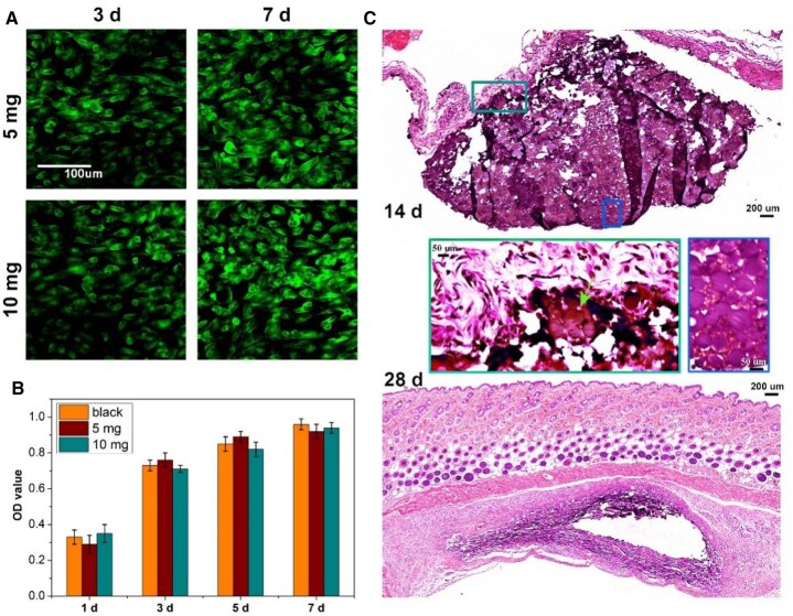
Figure 5.
Biocompatibility and degradability of CS-PPPDA&PPP-PA(10) microspheric gels. (A) Representative fluorescence images observed by live/dead staining assay to show the viability of fibroblasts co-cultured with the microspheric gels. (B) CCK-8 assay to demonstrate the cytotoxicity of microspheric gels in cell proliferation (n = 5). (C) H&E staining of subcutaneously implanted microspheric gels at 14- and 28-day post-implantation (n = 3).

