
FIGURE 6.
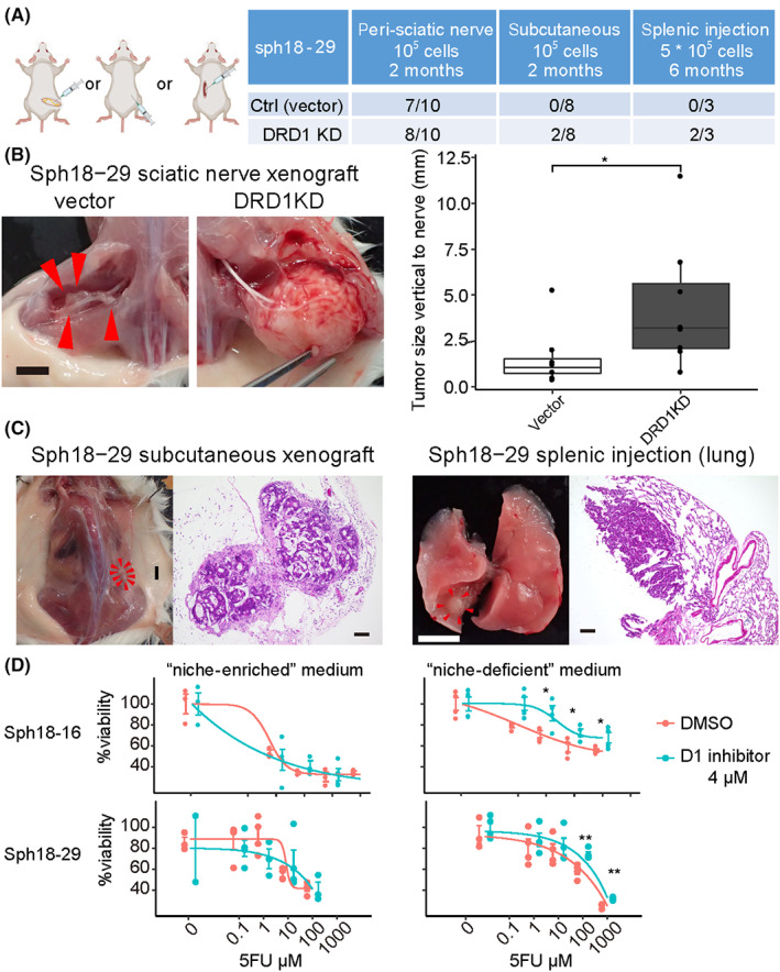
D1 inhibition enhancing cancer stem cell (CSC)‐related capacities in the absence of exogenous niches. (A) Peri‐sciatic nerve xenograft, subcutaneous xenograft, and splenic injection assay of Sph18‐29. Vector or two independent DRD1‐targeting shRNA is transduced to Sph18‐29. Table of the result (right). (B) Gross images of xenografted tumor in the peri‐sciatic nerve (left). Control patient‐derived cancer organoid (PDO tumor forming only along the nerves (arrowhead). Scale bar, 5 mm. The tumor size vertical to the nerve (mm) is measured (right). Wilcoxon rank‐sum test is used. (C) Macroscopic and HE‐stained histological images of a subcutaneously xenografted tumor and lung metastasis of DRD1‐KD sph18‐29. Scale bar, 5 mm in macroscopic images and 100 μm in histological images. (D) CCK8 proliferation assay of Sph18‐16, 29 with 5FU in the niche‐enriched or niche‐deficient medium, n = 4. The result of cisplatin and gemcitabine is shown in Figure S12C. Two‐tailed unpaired Student's t‐test is used at each concentration