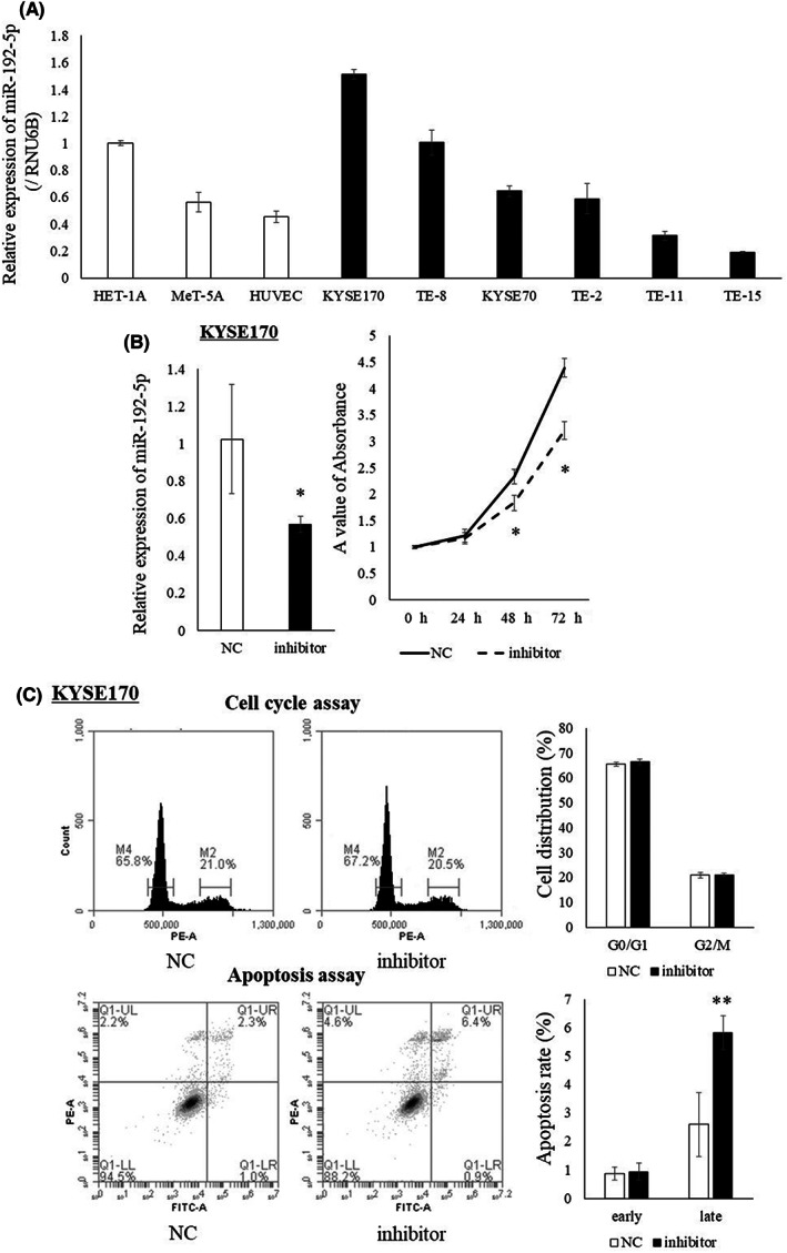
FIGURE 1.

(A) Expression of miR‐192‐5p in EC cell lines and normal cell lines. Expression of miR‐192‐5p was highest in KYSE170 cells and lowest in TE‐15 cells. (B) Expression of miR‐192‐5p in KYSE170 cells was downregulated to 60% using the miR‐192‐5p inhibitor. Proliferation ability of KYSE170 cells was significantly suppressed by the miR‐192‐5p inhibitor. (C) There was no change in cell cycle analysis by the miR‐192‐5p inhibitor. In apoptosis assay, the number of late apoptosis cells was increased by miR‐192‐5p inhibition. All experiments were performed in triplicate and results are shown as mean ± SD. Unpaired t‐test was used to analyze the data (*p < 0.05; **p < 0.01). NC, negative control.
