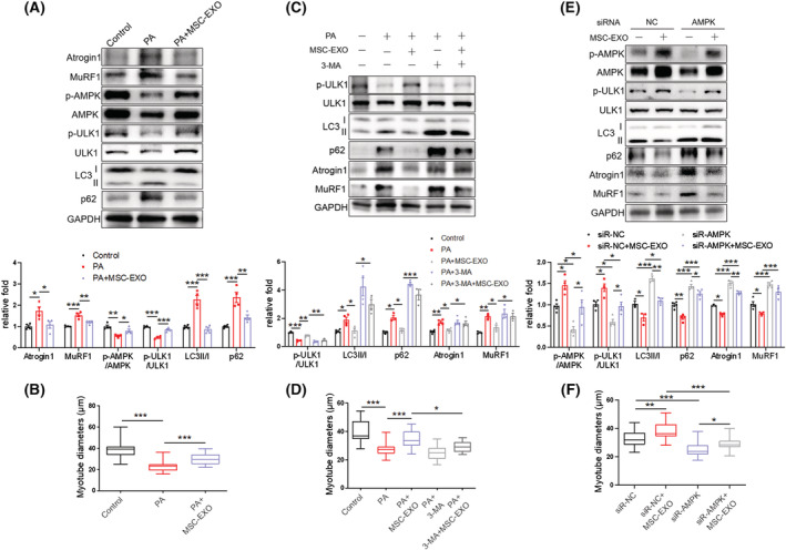
Figure 8.

MSC‐EXO counteracts PA‐induced myotube atrophy by enhancing the AMPK/autophagy signalling. (A) Western blot analysis of Atrogin 1 and MuRF1, p‐AMPK (T172) and p‐ULK1 (S555), LC3 and p62 in C2C12 myotubes treated with PA and MSC‐EXO (n = 4). (B) Diameters of C2C12 myotubes treated with PA and MSC‐EXO. (C) Western blot analysis of p‐ULK1(S555), LC3, and p62, Atrogin 1 and MuRF1 in C2C12 myotubes treated with MSC‐EXO and autophagy inhibitor 3‐MA (n = 3–4). (D) Diameters of C2C12 myotubes treated with MSC‐EXO and 3‐MA. (E) Western blot analysis of p‐AMPK (T172) and p‐ULK1 (S555), LC3 and p62, Atrogin1 and MuRF1 in C2C12 myotubes transfected with AMPK siRNA and treated with MSC‐EXO (n = 3–4). (F) Diameters of C2C12 myotubes transfected with AMPK siRNA and treated with MSC‐EXO. Quantification of bands was performed using ImageJ software. Data are mean ± SEM. (*P < 0.05, **P < 0.01, ***P < 0.001 using one‐way ANOVA followed by Tukey's test).
