Figure 5.

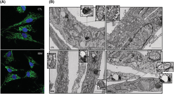
Mitochondrial morphology in IBM versus CTL fibroblasts. (A) Mitochondrial network in a representative image of IBM versus CTL fibroblasts obtained by confocal microscope (TOMM20 in green; nuclei in blue with DAPI, scale 10 μM) displaying reduced mitochondrial elongation in IBM (associated to poor mitochondrial health). (B) Transmission electron microscopy (TEM) images of IBM versus CTL fibroblasts. In IBM fibroblasts, some mitochondria lacked cristae (see inserts) presenting a discontinuous cristae distribution. Also, increased mitochondrial fission (see insert) could explain the smaller and more circular shape of these organelles associated with unhealthier mitochondria. Additionally, the presence of impaired autophagosome‐lysosome structures in IBM patients (see inserts) might be a result of incomplete autophagy. Briefly, these findings highlight a mitochondrial impairment in IBM fibroblasts. Magnification: 30 000×, scale 1 μm for each picture and 0.2 μm for inserts.
