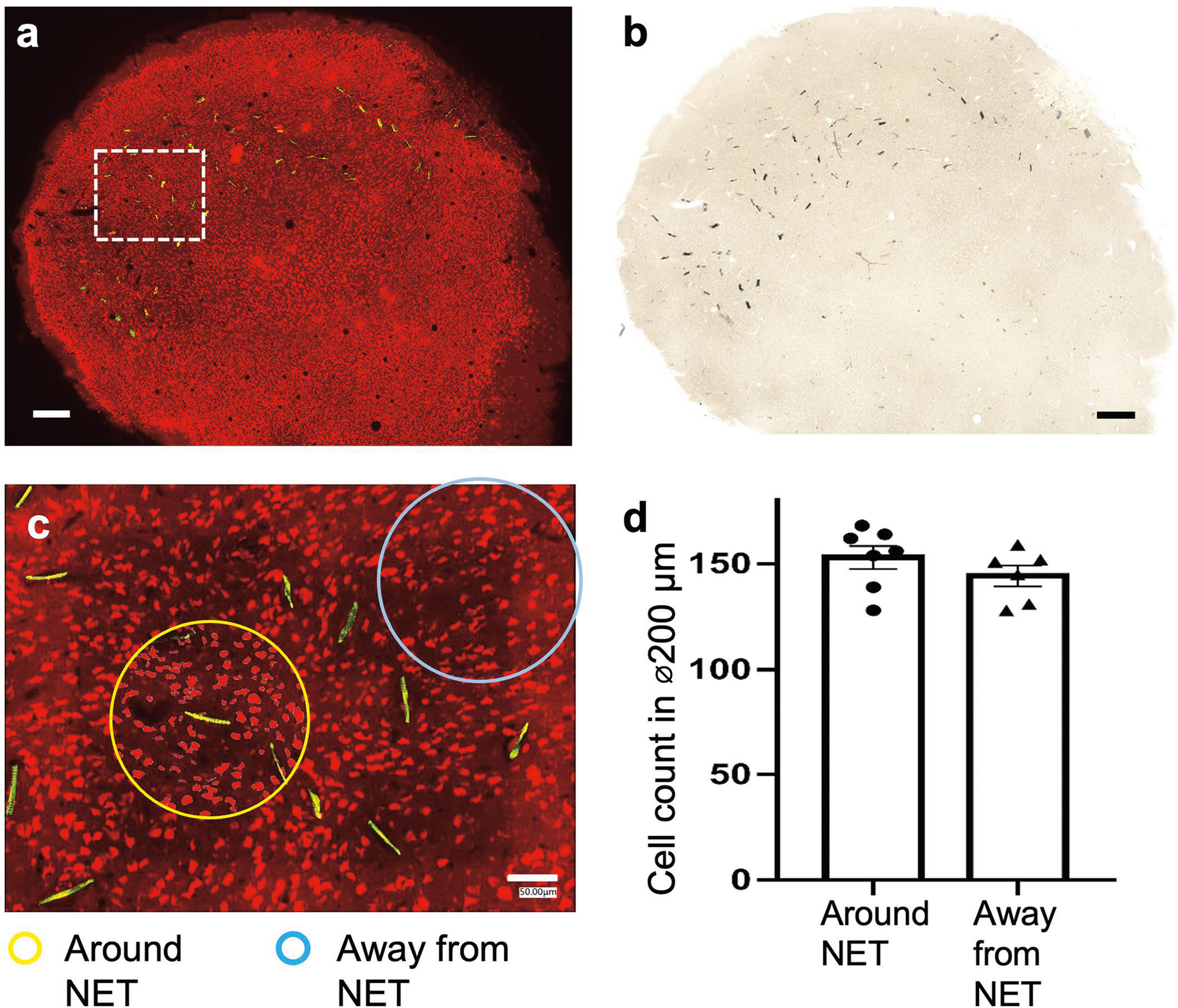
Extended Data Fig. 1 |. Immunohistochemistry analysis showing the tissue-NET interface at a high implantation density.

One mouse was implanted in visual cortex with 10 type-I NET modules, at inter-shank spacings of 150 μm and inter-module spacings of 250 μm. Fluorescent (a) and brightfield (b) microscopy show no observable scarring in the implanted regions. Neuron: red; NETs: yellow. (c), We counted numbers of neurons in seven randomly-chosen 200-μm-diameter regions centering around NETs (yellow circled regions as an example) and five 200-μm-diameter randomly-chosen regions away from NETs (cyan circled regions as an example). We found no significant difference between the two groups using unpaired T test, p = 0.275 (d), suggesting no significant neuronal loss induced by dense NET implants. These results are consistent with our prior studies using single or few NETs (10, 13). Scalebars: 250 μm (a and b) and 50 μm (c).
