Fig. 3.
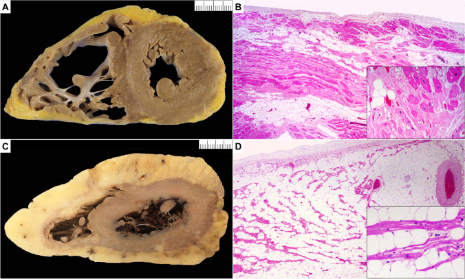
Arrhythmogenic cardiomyopathy (ACM) and pitfall (fatty heart). A Macroscopic short axis cut with fat infiltration from epicardial surface of the anterior, lateral, and posterior wall of the right ventricle with fat infiltration also of the lateral left ventricle wall. B Microscopic section shows fat admixed with collagen. There is individual myocyte and bundle degeneration (insert) in keeping with ACM (hematoxylin and eosin stain). C Macroscopic short axis cut with circumferential increase in epicardial fat surrounding both right and left ventricle. D Microscopic section shows mature fat admixed with myocyte bundles. There is no myocyte degeneration or increase in collagen (hematoxylin and eosin stain)

