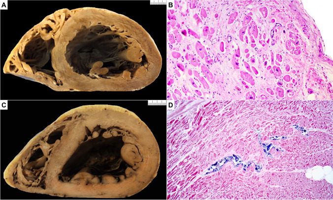
Fig. 4.
Dilated cardiomyopathy (DCM) and pitfall (postmortem putrefaction). A Macroscopic short axis cut with dilated left ventricle chamber and thinning of the left ventricular free wall. B Microscopic section shows interstitial and replacement collagen admixed with hypertrophied and degenerate myocytes in keeping with DCM (hematoxylin and eosin stain). C Macroscopic short axis cut with dilated left ventricle chamber and thinning of the left ventricular free wall. Note also the dark discoloration of both right and left ventricle. D Microscopic section shows hazy myocytes with separation of individual myocytes and myocyte bundles. There is bacterial proliferation within capillaries. Gas bubble formation is also noted (hematoxylin and eosin stain)

