Figure 2.
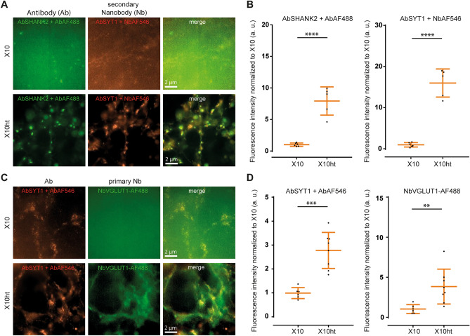
Applicability of nanobodies in X10ht, compared to homogenization with proteinase K (original X10 protocol). (A) Exemplary epifluorescence images of hippocampal cultured neurons immunostained with antibodies (Ab) for the postsynaptic protein SHANK2 and the presynaptic protein synaptotagmin1 (SYT1), revealed by secondary antibodies (AbSHANK2 + AbAF488) or secondary nanobodies (Nb) (AbSYT1 + NbAF546). (B) A pronounced increase in fluorescence intensity in both immunostainings is obtained when samples are autoclaved (X10ht) compared with the original X10 protocol. N = 6 images with several ROI analyzed for AbSHANK2 + AF488 X10 and AbSYT1 + NbAF546 X10, and N = 5 images with several ROI analyzed for AbSHANK2 + AF488 X10ht and AbSYT1 + NbAF546 X10ht. (C) Exemplary images of SYT1 antibody immunostainings, revealed by a secondary antibody (AbSYT1 + Ab AF546), in parallel with labeling for the vesicular glutamate transporter 1 (VGLUT1) with primary nanobodies coupled to AF488 (NbVGLUT1-AF488). (D) The quantification of the fluorescence signal revealed significantly increased intensities with X10ht. N = 6 images with several ROI analyzed for AbSYT1 + AbAF546 X10 and NbVGLUT1-AF488 X10, and 8 images with several ROI analyzed for AbSYT1 + AbAF546 X10ht and NbVGLUT1-AF488 X10ht from 2 gels. Data are presented as single data points, mean ± SD.

