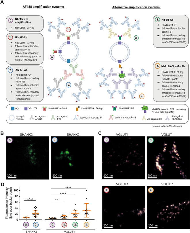
Figure 4.
Pre-expansion labeling of presynaptic markers with different signal amplification systems in X10ht. (A) Schematic visualization of the different amplification systems. See the main text for more details. Final used fluorophores are Alexa Fluor488 (AF488) or Abberior Star635P (AS635P). (B) Representative confocal images of expanded neurons immunostained with either the antibody-AF488-based amplification system for SHANK2, or (C) with the different nanobody-based amplification systems detecting VGLUT1. (D) Quantitative analysis of SHANK2 with N = 7 images for 0. Ab w/o amplification, and N = 22 images for 2. Ab-AF-Ab from 3 experiments; N = 18 images for 0. Nb w/o amplification (VGLUT1) and 1. Nb-AF-Ab, N = 13 images for 3. Nb-BT-Ab, and N = 14 images for 4. NbALFA-SpaMo-Ab all to detect VGLUT1, with several ROIs analyzed per image generated from 2 experiments. Data are presented as single data points, mean ± SD.

