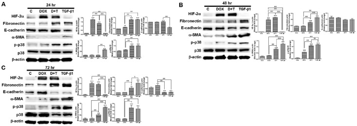
Fig. 4.
Effect of HIF-2α overexpression by DOX induction in renal tubular epithelial cells (TECs) under stimulation with transforming growth factor-β1 (TGF-β1) for 24, 48, and 72 hr. (A-C) HIF-2α, fibronectin, E-cadherin, α-SMA, and p-p38 protein levels were determined using western blotting of TECs treated with 10 ng/ml of TGF-β1 and 5 μg/ml of DOX for the indicated period (left panel). Band intensities were quantified via computerized densitometry (right panel). *P < 0.05 and ***P < 0.001 compared to the controls; #P < 0.05 and ##P < 0.01 between the two groups.

