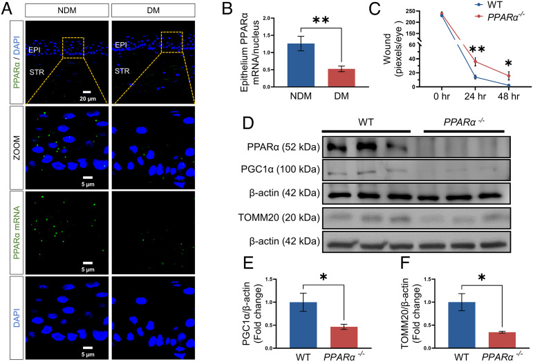
Fig. 2.
Decreased PPARα levels in diabetic human corneas. (A) Representative images of the PPARα mRNA (green) using RNAscope fluorescent assay, with the nuclei counterstained with DAPI (blue) in corneal sections from donors with NDM and DM. EPI, epithelium; STR, stroma. The scale bars represent 20 μm for the upper image and 5 μm for the bottom three images. (B) The total PPARα mRNA puncta in the corneal epithelium were counted by ImageJ and normalized by the number of nuclei to quantify the expression of the PPARα mRNA (n of NDM = 11, n of DM = 13). (C) Wound healing in the corneas from PPARα−/− and WT mice was quantified after fluorescein staining using the pixel per eye with ImageJ (n = 10). (D–F) Representative Western blots for PPARα, PGC-1α, TOMM20, and β-actin and densitometry quantification in the corneas from 5-mo-old PPARα−/− mice and WT littermates (n = 3). All values are mean ± SEM. *P < 0.05; **P < 0.01.

