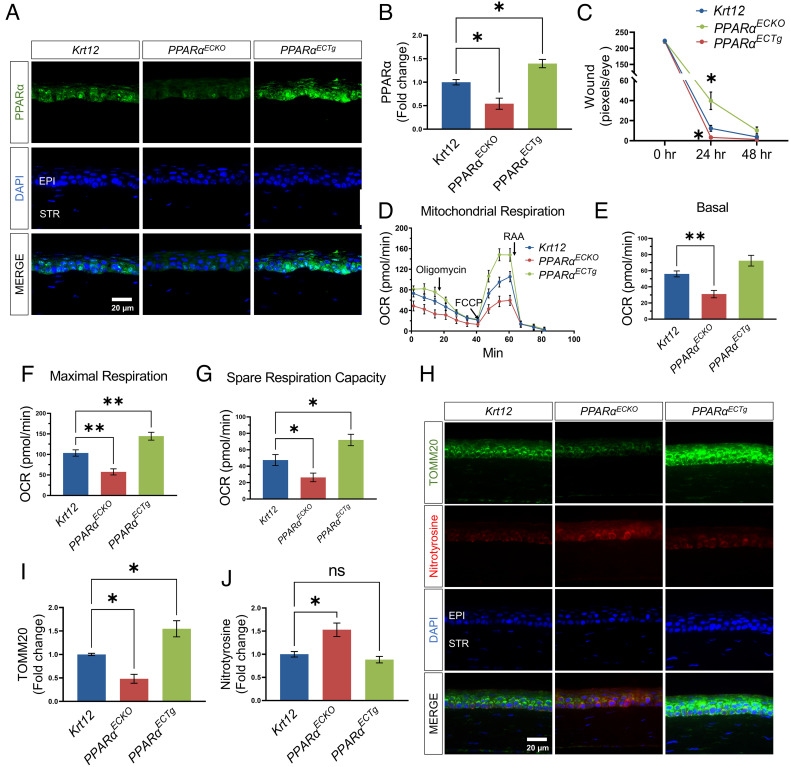
Fig. 4.
Corneal epithelium-specific PPARα KO impaired mitochondrial function and delayed corneal wound healing. (A) Representative immunohistochemistry images for PPARα staining (green) in the corneas from Krt12-Cre (Krt12)PPARαECKO, and PPARαECTg mice. (B) The intensity of PPARα in the epithelial layer in (A) was quantified using ImageJ (n = 3). (C) The corneal wound was quantified after fluorescein staining in Krt12-CrePPARαECKO and PPARαECTg mice and expressed by the pixel per eye with ImageJ (n = 12 to 14). (D–G) Mitochondrial stress test in the corneas from Krt12-CrePPARαECKO, and PPARαECTg mice (n = 10). (H) Representative immunohistochemistry images of TOMM20 (green) and nitrotyrosine (red) in the corneas from Krt12-CrePPARαECKO, and PPARαECTg mice. I&J: The intensities of TOMM20 (I) and nitrotyrosine (J) in the epithelial layer in (H) were quantified using ImageJ (n = 3). Values are mean ± SEM. *P < 0.05; **P < 0.01; ns, nonsignificant.

