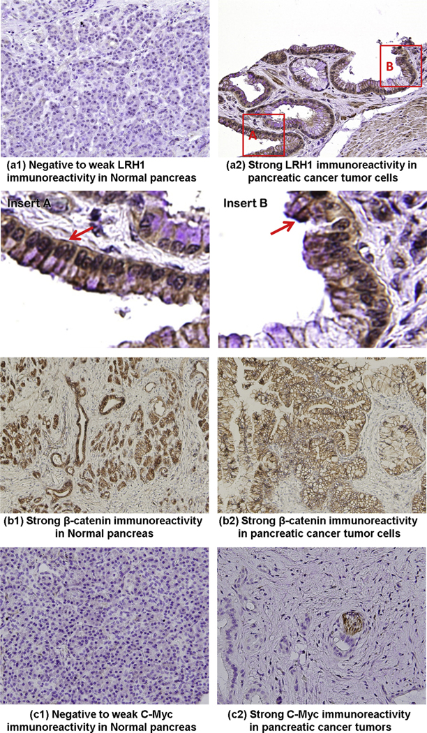
Fig. 6.

Expression of LRH1 and related genes in PC. LRH1 was overexpressed in tumor tissues (a2) compared with adjacent normal pancreas (a1) of patients with PC. Negative staining was observed for all components of normal exocrine pancreas. In tumor cells, an elevated level of LRH1 was detected either in the nuclei (insert A) or in the cytoplasm (insert B) or both. The original magnification (40×) is specified. The expression of β-catenin (b1–b2) was comparable between the tumorous and adjacent normal pancreas. Weak-negative staining for c-Myc (c1) was observed for all components of normal exocrine pancreas. In tumor cells, an elevated level of c-Myc (c2) was detected. The original magnification (40×) is specified.
