Fig. 2.
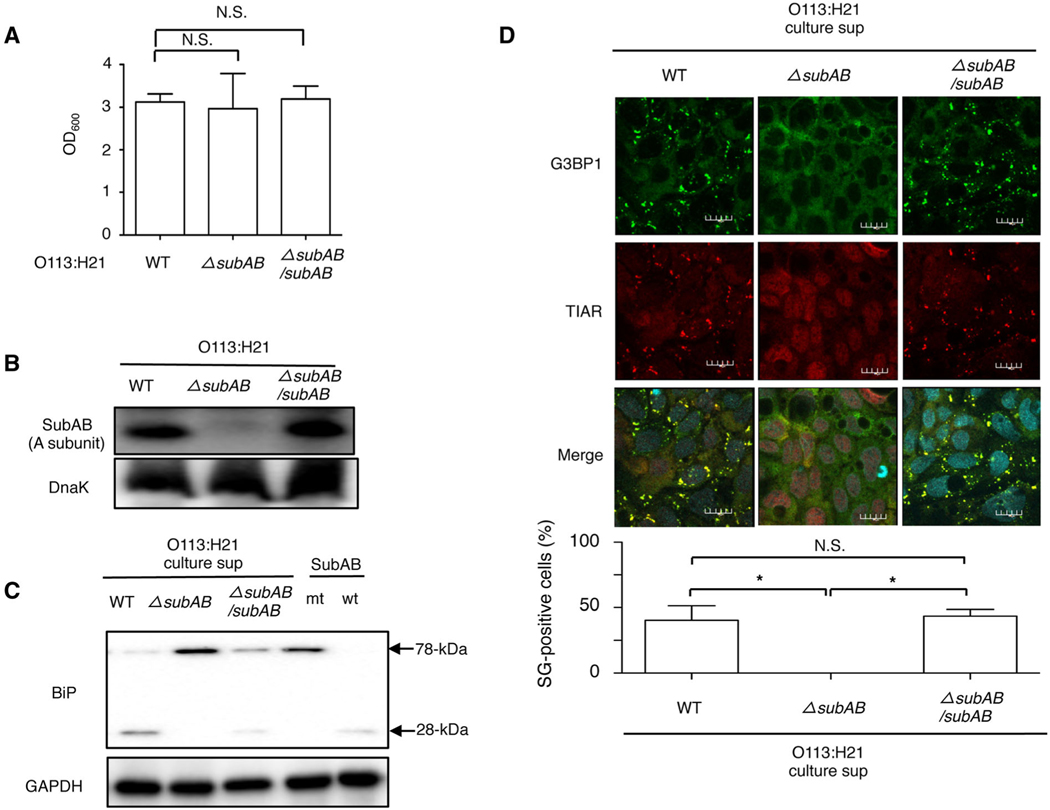
STEC O113:H21-produced SubAB induces SG formation.
A. Wild-type, ΔsubAB or ΔsubAB/subAB O113:H21 strains were cultured in BHI medium for 12 h at 37°C. Bacterial growth was measured by optical density at 600 nm (OD600).
B. The wild, ΔsubAB or ΔsubAB/subAB O113:H21 strains lysates were subjected to immunoblotting with anti-SubAB antibody or anti-DnaK antibody as a loading control.
C. Caco2 cells were incubated with purified SubAB (wt), SubAS272AB (mt), culture supernatant of wild-type, ΔsubAB or ΔsubAB/subAB O113:H21 strains for 1 h at 37°C. Cell lysates were subjected to immunoblotting with anti-BiP antibodies. GAPDH served as a loading control.
D. The culture supernatant of wild-type, ΔsubAB or ΔsubAB/subAB O113:H21 strains were added to Caco2 cells, which were incubated for 6 h, fixed with 4% PFA, reacted with anti-TIAR antibody (red) and anti-G3BP1 antibody (green) and observed by confocal microscopy. Cell nuclei were stained by DAPI (cyan). The rate of SG formation is presented as mean ± SD from four different fields, which included at least 20 cells/field (bottom panel). Bars represent 20 μm. All experiments were repeated two times with similar results, and significance is *P < 0.05.

