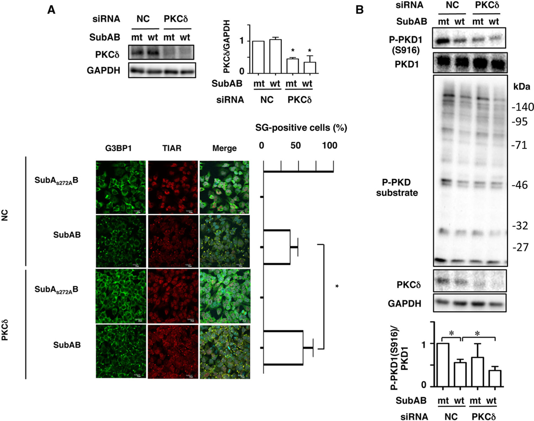
Fig. 6.
Protein kinase Cδ-knockdown enhances SubAB-induced SG formation.
A. Control (NC) or PKCδ siRNA-transfected cells were incubated with SubAS272AB (mt) or SubAB (wt) for 3–4 h. Cell lysates were subjected to immunoblotting with anti-PKCδ and anti-GAPDH antibodies. Quantification of the level of PKCδ/GAPDH was performed by densitometry (right panel). Data are means ± SD of values from three experiments, with n = 3 per experiment. Statistical significance is *P < 0.05. The fixed cells were reacted with the indicated antibodies and observed by confocal microscopy (lower panel). The rate of SG formation is presented as mean ± SD from five different fields, which included at least 20 cells/field (right panel). Bars represent 20 μm. Experiments were repeated three times with similar results, and significance is *P < 0.05.
B. Control (NC) and PKCδ siRNA-transfected cells were incubated with SubAS272AB (mt) or SubAB (wt) for 3–4 h. Cell lysates were subjected to immunoblotting with the indicated antibodies. GAPDH served as a loading control. Experiments were repeated three times with similar results. Quantification of the level of P-PKD1 (S916)/PKD1 was performed by densitometry (bottom panel). Data are means ± SD of values from three experiments, with n =3 per experiment. Statistical significance is *P < 0.05.

