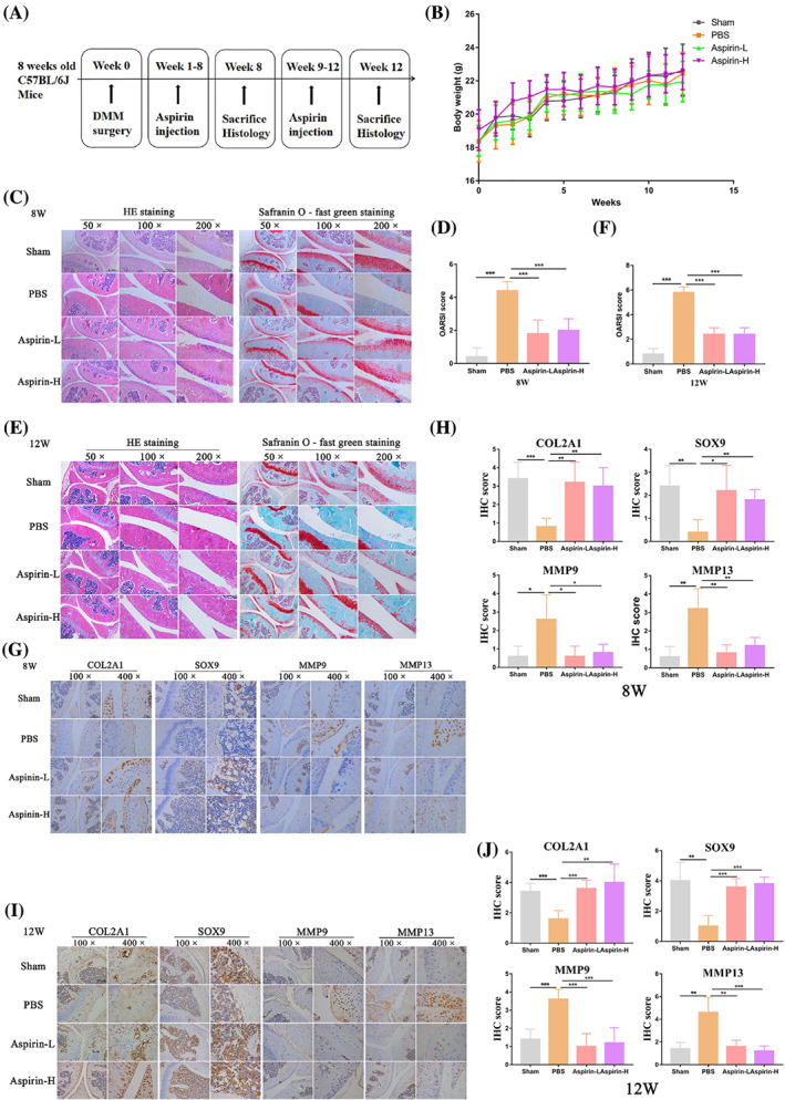
FIGURE 7.

Aspirin mitigates the progression of cartilage degeneration in a mouse DMM model. A schematic of the in vivo experiment. After DMM surgery, mice were injected with aspirin or PBS for 12 weeks (n = 10 per group). (B) The body weights of mice from different treatment groups were measured after 12 weeks. (C) Representative H&E and safranin O‐fast green staining images of the left knee joint of mice after exposure to different treatments for 8 weeks (n = 5 per group). (D) The severity of the OA‐like phenotype 8 weeks after DMM surgery in (C) was analysed by the Osteoarthritis Research Society International (OARSI) score system. (E) Representative H&E and safranin O‐fast green staining images of the left knee joint of mice after exposure to different treatments for 12 weeks (n = 5 per group). (F) The severity of the OA‐like phenotype 12 weeks after DMM surgery in (E) was analysed by the Osteoarthritis Research Society International (OARSI) score system. (G) IHC staining of chondrogenic markers (COL2A1 and SOX9) and catabolic markers (MMP9 and MMP13) in the left knee joint of mice after different treatments for 8 weeks. (H) IHC staining scores of (G). (I) IHC staining of chondrogenic markers (COL2A1 and SOX9) and catabolic markers (MMP9 and MMP13) in the left knee joint of mice after different treatments for 12 weeks. (J) IHC staining scores of (I). COL2A1, collagen type II alpha 1 chain; MMP9, matrix metallopeptidase 9; MMP13, matrix metallopeptidase 13; SOX9, SRY‐box transcription factor 9. *p < 0.05, **p < 0.01, ***p < 0.001. Scale bars: 0.5 mm (50× figures), 250 μm (100× figures), 100 μm (200× figures), 50 μm (400× figures)
