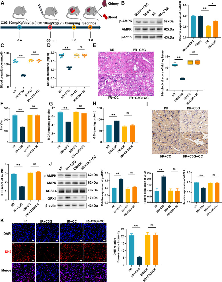
Fig. 7.
C3G alleviated I/R-AKI via activation of AMPK in vivo. A Schematic of renal I/R generation and treatment of C3G and CC at different period; B The protein level of activation of AMPK via C3G was analyzed by Western blot. (n = 3); C, D BUN and Scr levels in I/R-AKI mice. (n = 6); E Representative micrographs of HE staining of kidney from I/R, I/R + C3G, I/R + CC, I/R + C3G + CC group. The pathological scores of kidney injury were graded. Scale bars = 20 μm. (n = 6); F–H The content of iron, MDA, GSH were determined in kidney tissues of different groups. (n = 6). I Representative immunohistochemistry images of 4-HNE were captured respectively and analyzed. Scale bars = 20 μm. (n = 3); J Western blot results of AMPK, GPX4 and ACSL4 in kidney with or without CC. (n = 3); K Representative immunofluorescence images of ROS were captured and analyzed by the ratio of ROS positive cells/total cells. Scale bars = 20 μm.*p < 0.05, **p < 0.001. C3G cyanidin-3-glucoside, I/R-AKI ischemia/reperfusion-induced acute kidney injury, AMPK AMP-activated protein kinase, CC Compound C, BUN blood urea nitrogen, Scr serum creatinine, HE hematoxylin and eosin; MDA malondialdehyde; GSH glutathione, 4-HNE 4-hydroxynonenal, GPX4 glutathione peroxidase 4, ACSL4 acyl-CoA synthetase long chain family member 4, 4-HNE 4-hydroxynonenal, ROS reactive oxygen species

