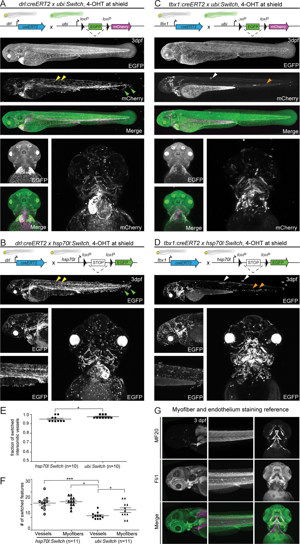
Figure 2. hsp70l:Switch high degrees of lineage labeling crossed to tissue-specific CreERT2 driver lines.

(A,B) drl:creERT2, and (C,D) tbx1:creERT2 crossed to ubi:Switch and hsp70l:Switch, respectively induced with 4-OHT at shield stage and imaged laterally and ventrally at 3 dpf. Quantifications depicted as bar diagrams with individual data points (E,F). Schematics of fluorophore cassettes for each Switch transgene are shown at the top of each panel and larval schematics represent secondary transgenic markers (A,B,C,D). drl:creERT2 yields more complete recombination when crossed to hsp70l:Switch (B) compared to ubi:Switch (A). The higher degree of recombination is detectable in the somitic myofiber (yellow arrowheads), and fin fibroblasts (green arrowhead) (A,B). A higher percentage of switched intersomitic vessels (ISV) is observed when combining drl:creERT2 with ubi:Switch than with hsp70l:Switch (n=10, unpaired t-test, p=0.0345) (E). tbx1:creERT2 yields less switching mosaicism when crossed to hsp70l:Switch (D), compared to ubi:Switch (C). Higher recombination efficiency is readily observed in trunk skin (white arrowhead) and hematopoietic cells (orange arrowheads) (C, D). When crossed to hsp70l:Switch, tbx1:creERT2 yields significantly more switched head vessels and myofibers compared to ubi:Switch (n=11, 1-way ANOVA p<0.0001) (F). Representative lateral and ventral images (A,B;D,E). (G) Max projections of z-stack confocal images of fli1a:EGFP transgenic zebrafish fixed at 3 dpf and stained for myofibers using an MF20 antibody to visualize vasculature and myofiber anatomy in the zebrafish head at 3 dpf.
