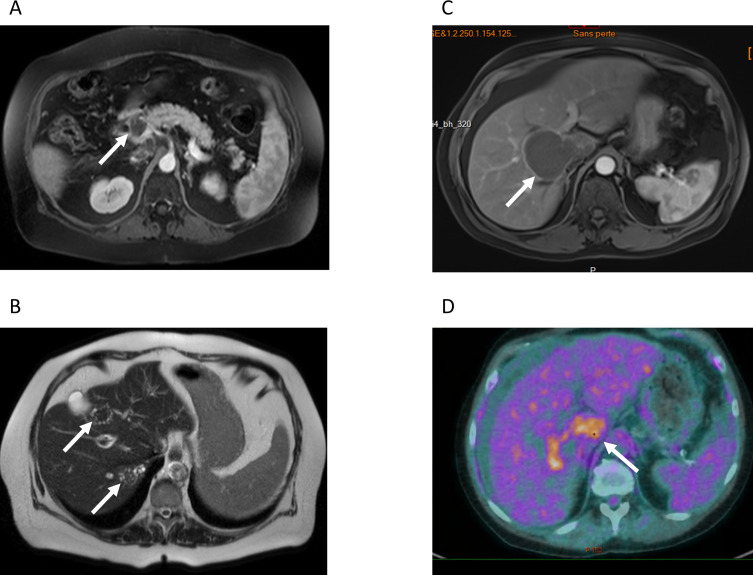
Figure 1.
Radiological findings. A) and B) MRI of patient #5 A) T1-weighted MRI of patient #5. White arrow: lesion in the head of the pancreas. This lesion was mistakenly interpreted as pancreatic cancer. B) T2-weighted, the white arrows point at the typical microcysts in T2-hypersignal. C) T1-weighted MRI of patient #4, showing a large well-delimited lesion tumor-like lesion, with invasion of the right portal branch D) PET-CT of patient #4, showing intense metabolic activity at the periphery of the lesion.

