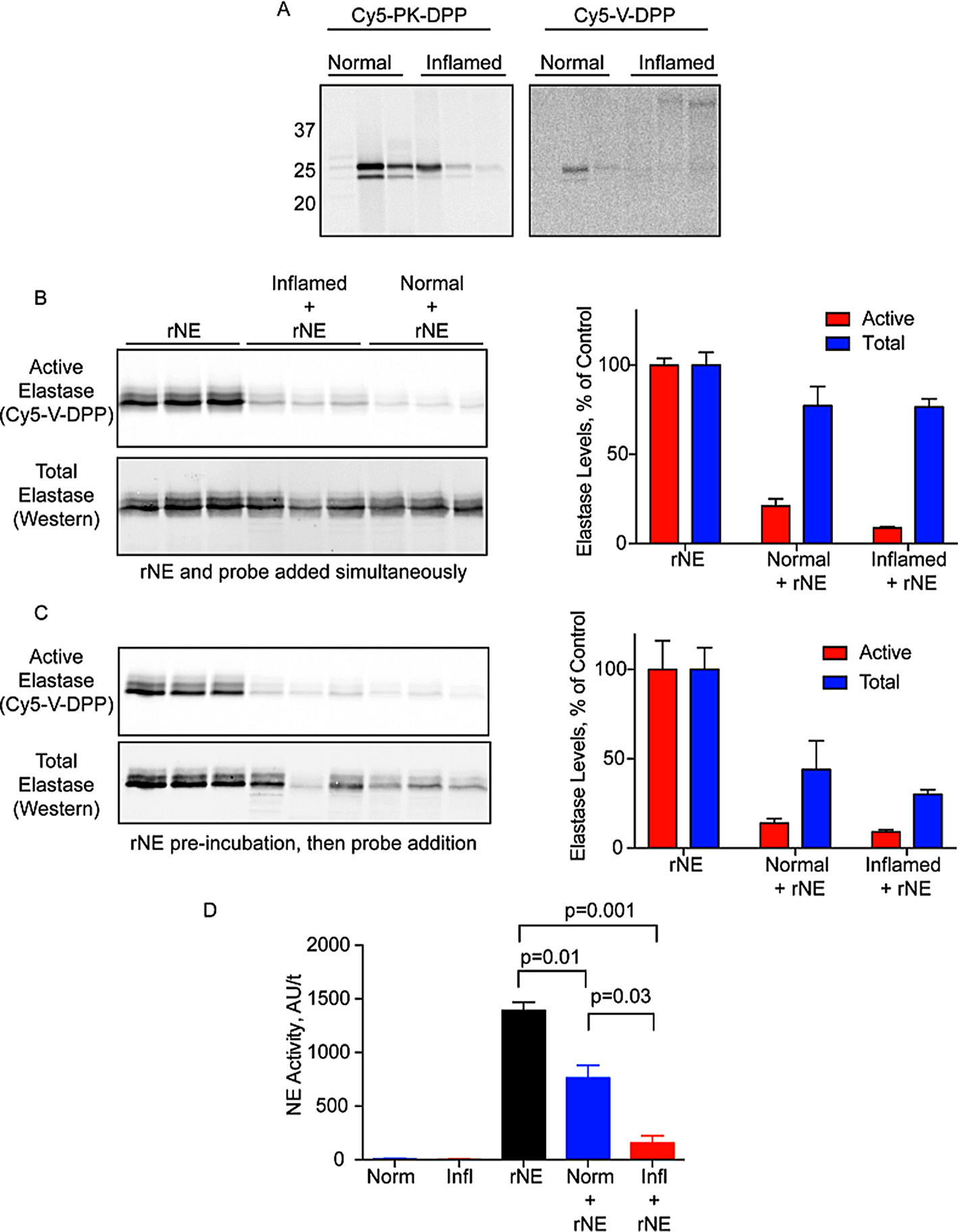
Fig. 5.

Ex vivo labeling of protease activation during acute colitis. A) Luminal fluids were collected from normal and inflamed colons and incubated with Cy5-PK-DPP or Cy5-V-DPP (1 μM). Proteins were resolved by SDS-PAGE and protease labeling was detected by measuring in-gel fluorescence. B) Recombinant neutrophil elastase was incubated with Cy5-V-DPP, either alone (rNE) or in the presence of luminal fluids (normal/inflamed + rNE). Elastase activity was then detected by in-gel fluorescence (top/red) followed by western blotting for total elastase (bottom/blue) Bands were quantified by densitometry. (Activity: rNE v Norm p = 0.0001; rNE v Infl p = 1.7e-5; Norm v Infl p = 0.03. Total: rNE v Norm p = 0.15; rNE v Infl p = 0.05; Norm v Infl p = 0.96. Norm Act v Tot p = 0.008; Infl Act v Tot p = 0.0001). C) Samples were treated as in B except rNE was preinbuated with the samples for 10 min prior to Cy5-V-DPP addition. (Activity: rNE v Norm p = 0.006; rNE v Infl p = 0.005; Norm v Infl p = 0.16. Total: rNE v Norm p = 0.05; rNE v Infl p = 0.005; Norm v Infl p = 0.43. Norm Act v Tot p = 0.13; Infl Act v Tot p = 0.002). D) Measurement of NE activity in luminal fluids of normal and inflamed mice in the presence and absence of rNE using the fluorogenic substrate probe AAPV-AMC (n = 3 mice per condition).
