Figure 1.
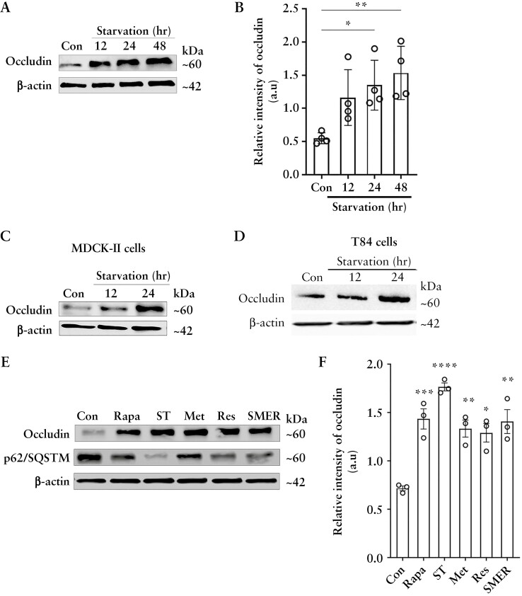
Autophagy enhances cellular occludin levels and enhances the TJ barrier against macromolecular flux. [A] Western blot for Caco-2 occludin levels after starvation. β-Actin is shown as a loading control. [B] Quantification of occludin in panel A using ImageJ software. Blot and densitometry data representative of more than three independent experiments. [C, D] Starvation increased occludin levels in MDCK-II and T84 cells. Blots are representative of three independent experiments. [E] Occludin levels from Caco-2 cells treated with rapamycin [Rapa, 500 nM], EBSS [ST], metformin [MET, 100 µM], resveratrol [RES, 100 µM] and small-molecule enhancer 28 [SMER, 50 µM]. SQSTM/p62 protein degradation indicates autophagy induction. Occludin levels were increased by all the autophagy inducers. [F] Quantification of occludin levels in panel E. Blot and densitometry analysis from three independent experiments. Student’s t-test or one-way ANOVA with Tukey’s post-hoc test. *, p < 0.05; **, p < 0.01; ***, p < 0.001; ****, p < 0.0001. a.u = arbitrary units.

