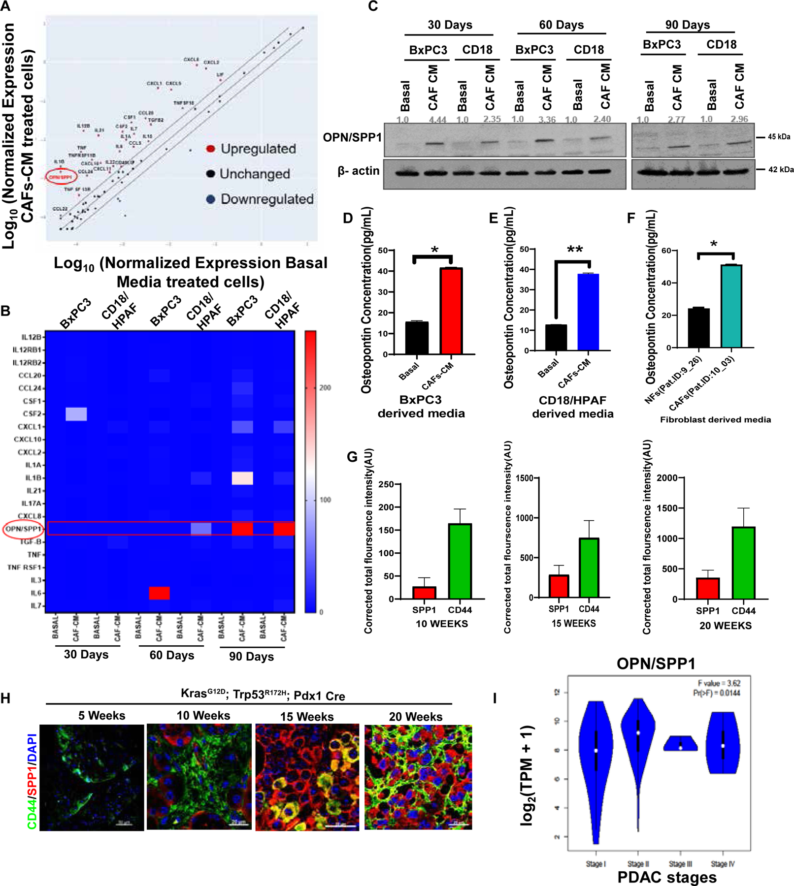
Figure 6. CAF-CM subsidizes the up-regulation of OPN/SPP1 in PC cells.

(A) PCR array for receptors and ligands molecules associated with stemness signaling from CAF-CM/Basal-exposed PC cells. (B) Heatmap of top differentially expressed genes of PCR array (cut off < 5 fold). (C) Immunoblotting of OPN/SPP1 protein expression in 30, 60, and 90-dayCAF-CM/Basal-exposed PC cells. β-actin was used as a housekeeping control. (D-F) The OPN/SPP1levels in culture media. Bar diagrams depict the OPN/SPP1levels in pg/mL in the serum-free culture media of NFs/CAF-CM-exposed cells. (G-H) Representative bar graphs and images are showing immunofluorescence staining of CD44 (green) and OPN/SPP1 (red) in 5, 10, 15, and 20 weeks old KPC (KrasG12D; p53R172H; Pdx1Cre) mice tissues. Nuclei were stained in DAPI (blue). n=3; Scale Bar= 20um. (I) Representative violin plot showing the OPN/SPP1 expressions on different stages of PAAD; n (T) =179; n (N) =171. Data represent mean ± SD (P values were calculated by Student t-test) *P <0 .05, **P <0 .01, ***P < 0.001.
