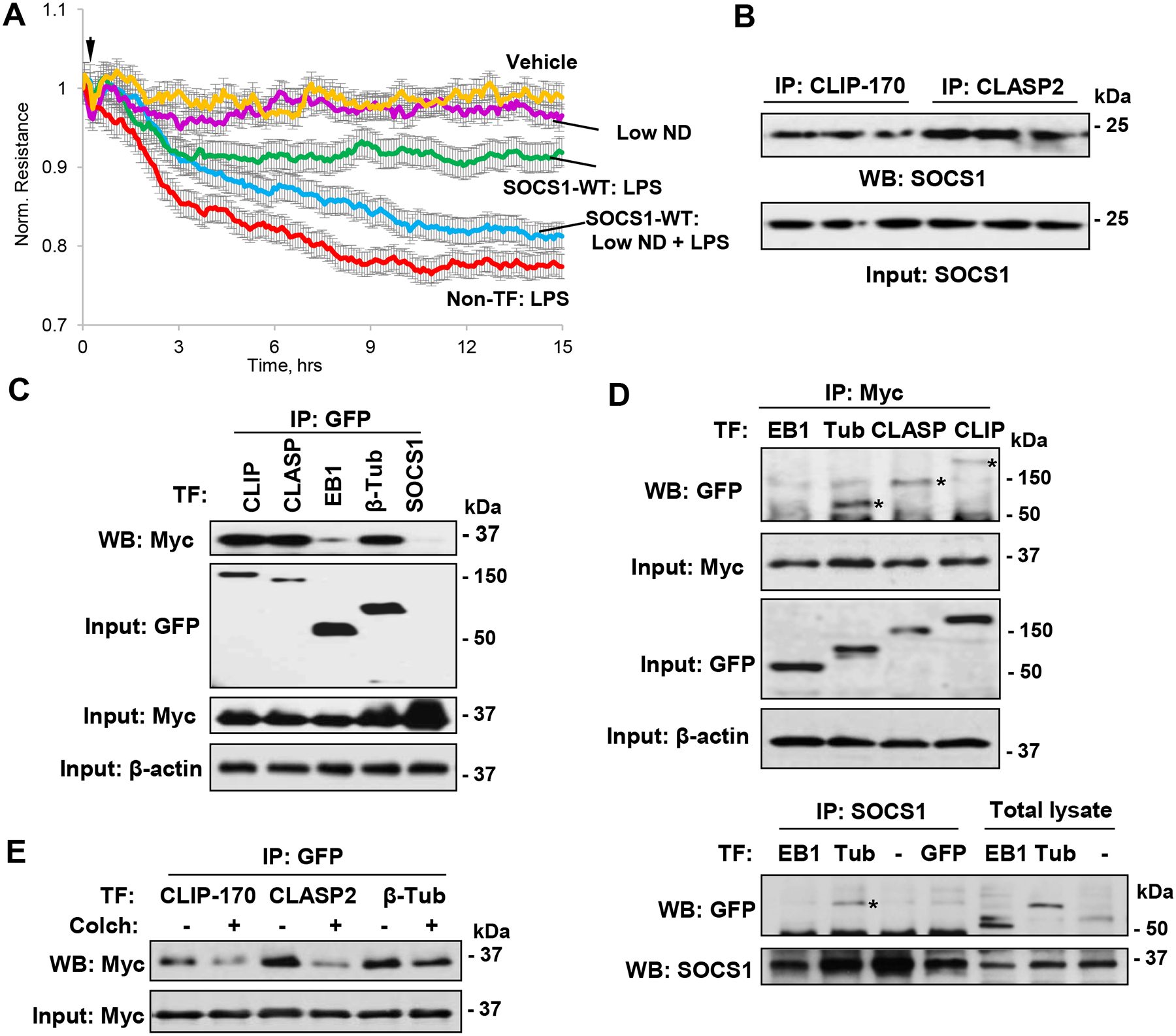
Figure 5. SOCS1 function on EC depends on MT.

A – SOCS1-WT expressing EC were exposed to LPS in the presence or absence of low dose of nocodazole (0.05 nM) and EC permeability was determined by monitoring TER over indicated time period. B – Co-IP assays with CLIP-170 and CLASP2 antibodies, followed by immunoblotting with SOCS1 antibody. C – Pulmonary EC co-transfected with GFP-tagged CLIP-170, CLASP2, EB1 or β-tubulin and Myc-SOCS1 were tested in co-IP assay with GFP antibody followed by immunoblotting with Myc-antibody. Probing of total cell lysates with GFP and Myc antibodies was used to verify ectopic protein expression; membrane re-probing for β-actin was used to ensure equal sample loadings. D – Cells were co-transfected with GFP-tagged EB1, β-tubulin, CLASP2 or CLIP-170 and Myc-SOCS1 plasmids. Upper panels: Co-IP assay with Myc antibody. Western blot analysis of immunocomplexes with GFP antibody was performed to detect co-immunoprecipitated proteins (marked by asterisks). Probing of total lysates for GFP and Myc was used to verify successful overexpression, and β-actin was used as a loading control. Lower panels: Co-IP assay with SOCS1 antibody. E – Cells with co-expression of GFP-tagged CLIP-170, CLASP2 or β-tubulin and Myc-SOCS1 for 24 h were stimulated with colchicine (0.5 μM, 1 h), followed by co-IP with GFP and immunoblotting with Myc antibody. Whole cell lysates were probed for Myc to verify ectopic SOCS1 expression.
