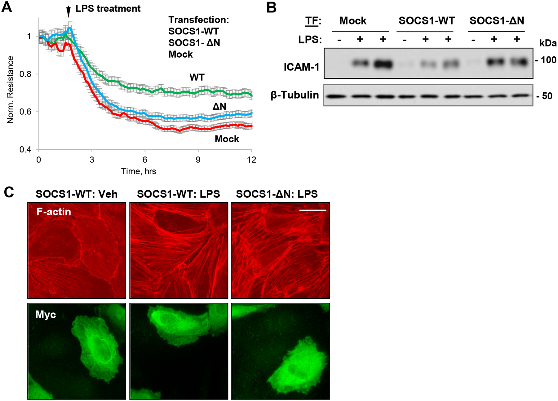
Figure 7. N-terminal region of SOCS1 is indispensable for its barrier protective and anti-inflammatory effects on EC.

HPAEC expressing SOCS1-WT or its ΔN mutant were stimulated with LPS (100 ng/ml). A – Permeability changes were monitored by TER measurements over time. B – Western blot analysis of ICAM-1 protein levels in control and LPS-stimulated (6 h) cells; β-tubulin was used as a loading control. C – Cells were treated with LPS, and immunofluorescence staining was performed with Texas Red phalloidin to visualize actin cytoskeleton. Co-staining with Myc antibody was performed to visualize transfected cells. Bar: 10 μm.
