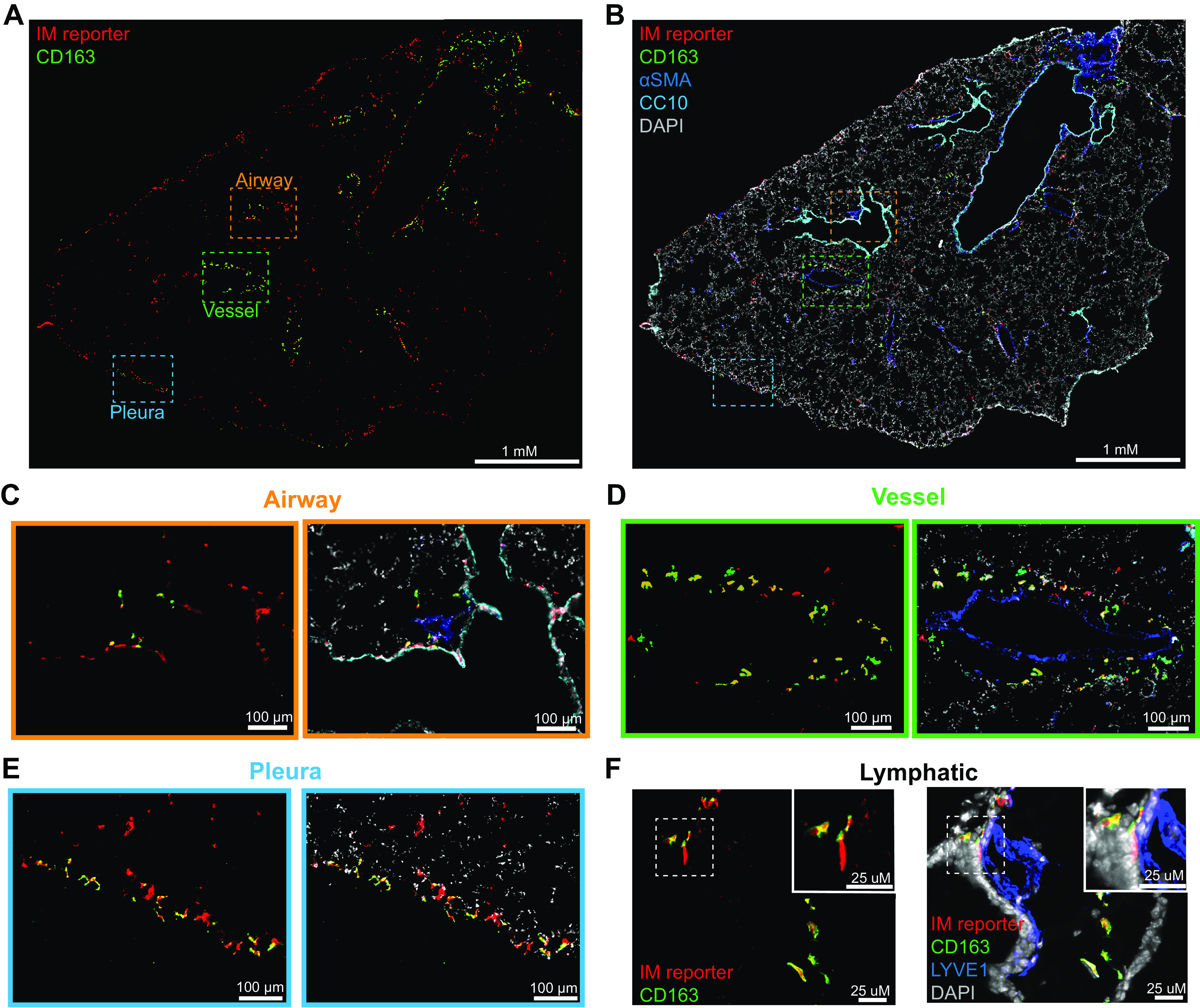
Figure 4.

CD163+ IMs are located near vessels, lymphatics, and pleura. CD163− IMs are adjacent to airways, alveoli and pleura. Immunofluorescence microscopy was performed on 10-μm thick frozen sections from male and female pulse-wait resident IM reporter mice (CX3CR1ERT2-Cre x R26Stop(fl/fl)tdTomato) at homeostasis (male mouse shown). CD163 was used to identify FRβ+ IMs. A: whole lobe projection of reporter positive IMs (red) and CD163 (green) imaged at ×20 on scanning microscope from a male mouse. B: airways (CC10/cyan) and vessels (αSMA/blue) and nuclei (DAPI/gray) are highlighted on whole lobe projection. Zoomed projection of IMs near an airway (C), a vessel (D), and the pleura (E). F: maximal intensity projection of 1 μM thick images of CD163 (green) IMs (red) and lymphatics (LYVE1/blue) using confocal microscopy. αSMA, α-smooth muscle actin; CC10, Clara Cell 10 kDa Protein; DAPI, 4′,6-diamidino-2-phenylindole; FRβ, folate receptor β; IM, interstitial macrophage.
