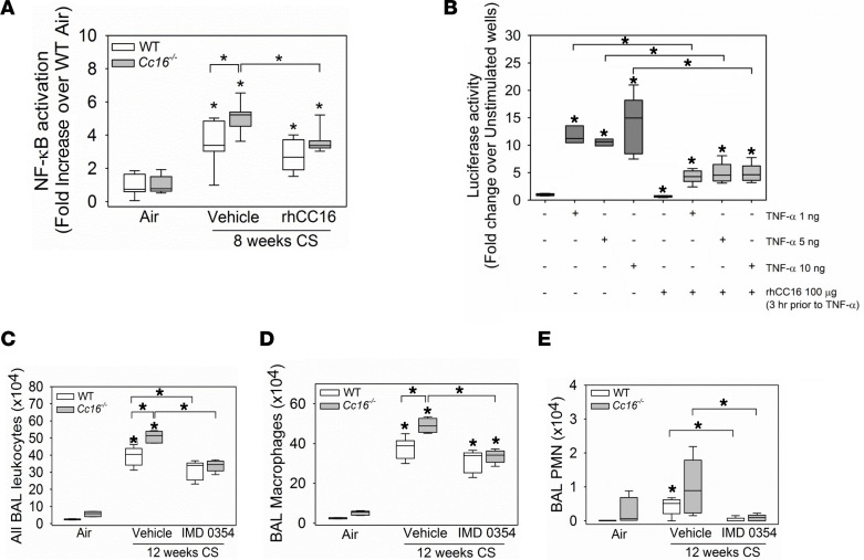
Figure 10. Treating CS-exposed Cc16–/– mice with rhCC16 reduces NF-κB activation in their lungs, and increased NF-κB activation in the lungs of CS-exposed Cc16–/– mice contributes to their exaggerated pulmonary inflammatory response to CS.
In A, WT and Cc16–/– mice were exposed to air (6 mice/group) or CS for 8 weeks (6–7 mice/group), and CS-exposed mice were treated thrice weekly with rhCC16 (75 μg of rhCC16) or vehicle. NF-κB that translocated to the nucleus was quantified in nuclear extracts of whole lung samples using a TransAM NF-κB kit. In B, NF-κB luciferase reporter A549 cells were grown to at least 80% confluence and preincubated for 3 hours with 100 μg/mL of rhCC16. Cells were then activated with rhTNF-α (1–10 ng/mL), and 8 hours later, the cells were lysed and luciferase activity was measured. In C, WT and Cc16–/– mice were exposed to air (3–5 mice/group) or CS for 12 weeks (10–12 mice/group), and CS-exposed mice were treated once daily with a solution of IMD0354 (6 mg/kg body weight) or vehicle via the intraperitoneal route during the last 6 weeks of the CS exposures (5–6 mice/group). BAL was performed, and BAL total leukocytes (C), macrophages (D), and PMNs (E) were counted. Boxes in the box plots show the medians and 25th and 75th percentiles, and whiskers show the 10th and 90th percentiles. Data were analyzed using 1-way ANOVAs followed by pairwise testing with Mann-Whitney U tests. In A and C–E, asterisks indicate P < 0.05 vs. air-exposed mice belonging to the same genotype or the group indicated; in B, asterisks indicate P < 0.05 vs. negative control in lane 1 or the group indicated.

