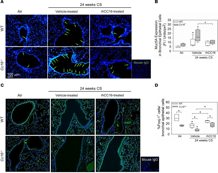
Figure 4. Treatment of mice with rhCC16 limits the development of mucus cell metaplasia and rescues Foxj1 expression in mice exposed chronically to CS.
WT and Cc16–/– mice were exposed to air (3–5 mice/group) or CS for 24 weeks (10–13 mice/group), and rhCC16 (75 μg of rhCC16; 5–7 mice/group) or vehicle (5–6 mice/group) was delivered thrice weekly by the i.n. route to CS-exposed mice for the last 12 weeks of the CS exposures. Lung sections were immunostained for Muc5ac (A and B) and Foxj1 (C and D), and the percentage of positively stained airway epithelial cells was determined. Arrows point to Muc5ac-positive cells. The box plots show the medians and 25th and 75th percentiles, and whiskers show the 10th and 90th percentiles. Data were analyzed using 1-way ANOVAs followed by pairwise testing with Mann-Whitney U tests. Asterisks indicate P < 0.05 vs. air-exposed mice belonging to the same genotype or the group indicated. FI, fluorescence Intensity; px, pixel.

