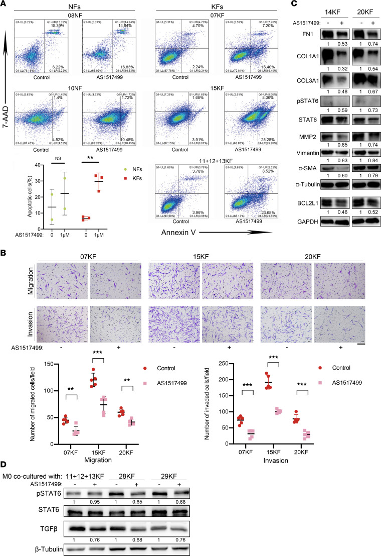
Figure 7. Inactivation of p-STAT6 with AS1517499 inhibited profibrotic activities.
(A) Annexin V/7-AAD double staining assay of NFs and KFs treated with or without AS1517499. NFs and KFs were treated with or without 1 μM AS1517499 for 16 hours and then subjected to Annexin V/7-AAD staining. Quantification of Annexin V positivity, representing apoptotic rates, is shown below. (B) Migration and invasion of KFs with or without AS1517499 treatment. KFs were treated with or without 500 nM AS1517499 for 24 hours and then analyzed by Transwell assay. Scale bar: 50 μm. Quantification of migrated and invaded cells is shown below. (C) Expression of mesenchymal markers (FN1, vimentin), collagen (COL3A1, COL1A1), MMP2, BCL2L1, α-SMA, p-STAT6, and STAT6 in KFs upon AS1517499 treatment. KFs were treated with or without 500 nM AS1517499 for 24 hours and then analyzed by Western blotting assay. (D) Expression of p-STAT6, STAT6, and TGF-β in THP-1 cells when cocultured with pretreated KFs (800 nM AS1517499 for 48 hours or negative control). Relative gray scales of p-STAT6/STAT6 and indicated protein/GAPDH are shown below the blots. Error bars represent SD. Experiments were performed at least 3 times. **P < 0.01, ***P < 0.001 by 2-tailed Student’s t test. NS, not significant (P > 0.05).

