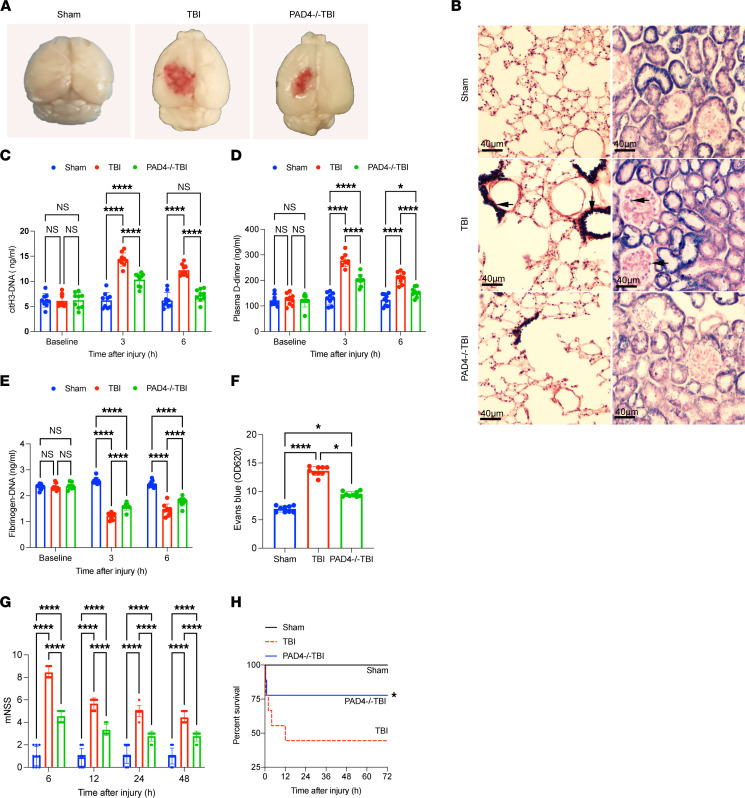
Figure 6. NETs induce coagulopathy in a TBI mouse model.
To evaluate the potential role of NETs in coagulopathy, we established a TBI mouse model. (A) Representative topical views of brains from each group (sham, TBI, and PAD4–/– TBI). (B) PTAH staining of the lungs and kidneys from each group. Fibrin deposition (black, arrow) in the microvasculature of the lungs and kidney was observed in TBI mice but not in the same organs of PAD4–/– mice. Scale bars: 40 μm. Plasma levels of citH3-DNA (C), fibrinogen (D), and D-dimer (E) in sham (n = 9), TBI (n = 9), and PAD4–/– TBI mice (n = 9). (F) Levels (OD unit) of Evans blue in the supernatants of tissue homogenates from mice in each group (n = 9). (G) Modified Neurological Severity Score (mNSS) (n = 9) of sham, TBI, and PAD4–/– TBI mice. (H) Kaplan-Meier survival analysis of sham, TBI, and PAD4–/– TBI mice. *P < 0.05 versus TBI mice. Data are presented as the mean ± SD. *P < 0.05; ****P < 0.0001 by 2-way ANOVA with Tukey’s multiple-comparison test (C–E and G) or Kruskal-Wallis test with Dunn’s multiple-comparisons test (F).

