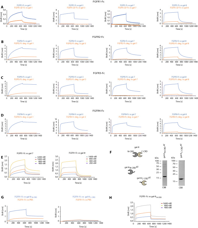
Fig. 3.
Galectins recognize the N-glycans of FGFRs. A–D BLI analyses of the interaction between N-glycosylated and de-glycosylated FGFRs, FGFs, and galectins. FGFR1-Fc (A), FGFR2-Fc (B), FGFR3-Fc (C), and FGFR4-Fc (D) were immobilized on Protein-A biosensors (blue lines) in a pairwise fashion with equimolar concentrations of the N-glycosylation-deficient mutant of FGFR1 (FGFR1.GF-Fc) or FGFR2-Fc, FGFR3-Fc, and FGFR4-Fc treated with PNGase F (red lines) and incubated with recombinant galectins to record the association and dissociation phases. Representative results from at least three independent experiments are shown. E BLI measurements of the kinetics of interaction between FGFR1-Fc, galectin-7 and -8. Kinetics values are provided in Table 1. F CBB stained gels of recombinant single CRD variants of galectin-8. G BLI binding curves of FGFR1-Fc with gal8N-CRD and gal8C-CRD variants. H BLI measurements of the kinetics of interaction between FGFR1-Fc and gal8N-CRD; binding parameters are provided in Table 1

