Figure 1:
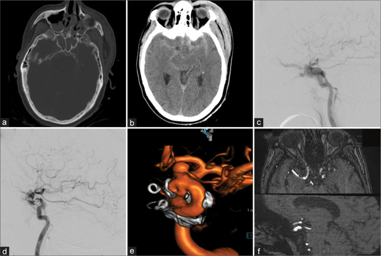
Patient 1, 55 F, traumatic brain injury. Admission computed tomography-head (a and b). Type A carotid-cavernous fistula in lateral diagnostic cerebral angiogram (DCA)-projection, the retrograde filling of the left superior ophthalmic vein is evident (c). Transvenous coil-embolization was performed in the acute setting (d). Follow-up DCA after 8 months showed a dorsal-medial focal outpouching of the internal carotid artery (ICA) as well as a prominent intracranial proper ophthalmic artery aneurysm (e). Flow diversion using Pipeline embolization device was performed and 6-month follow-up magnetic resonance angiography shows resolution of the ICA defect (f).

