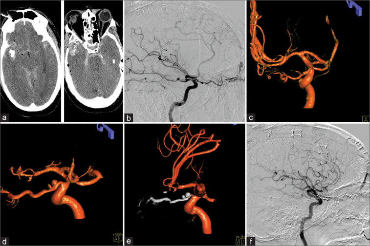
Figure 2:
Patient 2, 58 M, right temporal gunshot wound. Admission computed tomography-head (a). A traumatic right-sided ethmoidal dural arteriovenous fistula (dAVF) was evident on lateral diagnostic cerebral angiogram (b). Two minimal outpouchings of the intracranial and intracanalicular segment of the ophthalmic artery were identified as well (c). Three weeks after a complicated course, both proper ophthalmic artery aneurysm (POAAs) grew (d). Treatment of dAVF as well as POAAs was performed through Onyx embolization (e) with successful resolution of the POAAs as well as dAVF (f).

