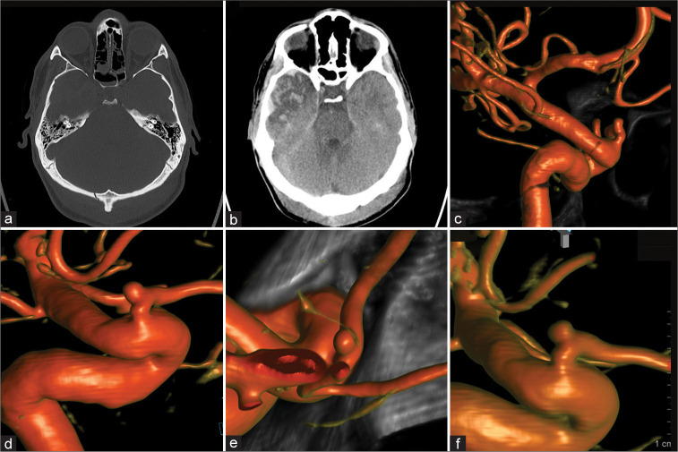
Figure 3:
Patient 3, 31 M, traumatic brain injury. Admission computed tomography-head (a and b). Four-month follow-up diagnostic cerebral angiogram (DCA) for potential vascular compromise was performed revealing a small intracranial proper ophthalmic artery aneurysm (c). Subsequent 4, 9, and 21 months DCA follow-ups demonstrated stable aneurysm size and morphology (d-f, respectively).

