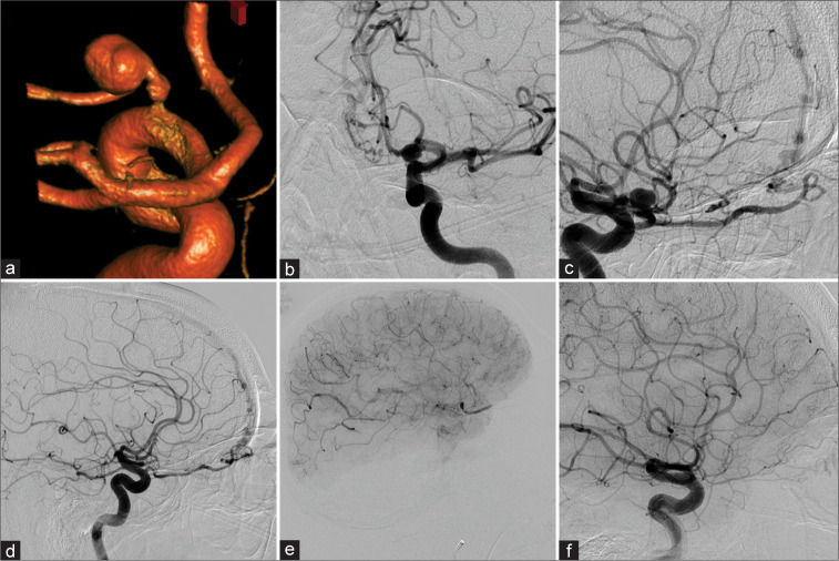
Figure 4:
Patient 4 was diagnosed with a left-sided proper ophthalmic artery aneurysm (POAA) (a-c) in a setting of a left (b and c) and right (d) sided ethomoidal dural arterio-venous fistula. After right-sided N-butyl cyanoacrylate (NBCA) embolization, the left side was treated with NBCA as well. However, the ophthalmic artery (OphA) demonstrated contrast stasis in the intraorbital segment – as well as in the POAA – in the postembolization run (e). Follow-up diagnostic cerebral angiogram (DCA) 5 months later revealed reconstitution of a small calibered OphA. However, vision did not recover beyond light-dark perception. Twelve years later, the DCA demonstrated equal findings (f).

