Figure 1:

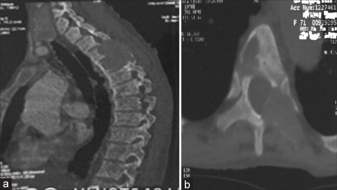
Computed tomography of the thoracic spine sagittal (a) and axial (b) images showing an osteolytic lesion regarding T3-T4-T5 with endocanalar extension.

Computed tomography of the thoracic spine sagittal (a) and axial (b) images showing an osteolytic lesion regarding T3-T4-T5 with endocanalar extension.