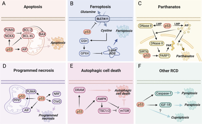
Fig. 1. The representative mechanisms for p53-mediated regulated cell death.
A p53 plays a key role in mitochondrial apoptosis. p53 transactivates the expression of the pro-apoptotic BCL-2 members, such as PUMA and NOXA, to inactivate anti-apoptotic BCL-2 and BCL-XL, resulting in the activation of the pore-forming protein, BAX, and the consequent release of numerous apoptogenic factors [86], such as AIF that is also encoded by a p53-target gene [52]. B p53 can either promote or inhibit ferroptosis in the context of different cancers [87]. One of the well-characterized mechanisms is that p53 represses the expression of SLC7A11, reducing GSH biogenesis, increasing lipid ROS, and thus promoting ferroptosis [9]. C Parthanatos is dependent on PARP1 activation, which leads to generation of PAR, nuclear translocation of AIF, and chromatinolysis [88]. p53 promotes LMP to facilitate nuclear translocation of DNase II with the help of AIF. DNase II then cleaves the DNA to activate the PARP1-AIF axis [54]. Also, p53, together with SIRT6, can directly activate PARP1 to induce parthanatos [89]. D Programmed necrosis is executed via both RIPK-dependent and independent pathways [90]. p53 promotes programmed necrosis by inducing the expression of AIF [91, 92], PUMA [93], CtsQ [94], and the long noncoding RNA NRF [95], or physically interacting with PPIF [96]. E Persistent activation of autophagy results in autophagic cell death. p53 activates AMPK signaling [36, 37] or DRAM expression [97, 98] to enhance this type of RCD. F p53 may also promote pyroptosis and paraptosis by regulating Caspase-1 [99] and IGF-1R [100], respectively.

