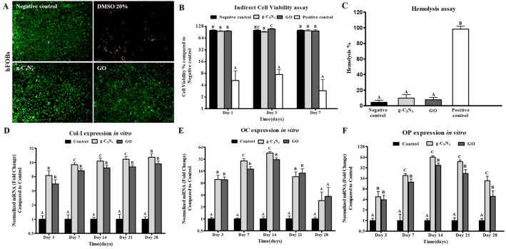
Figure 2.
In vitro experiments of g-C3N4 and GO. (A) Cell viability using Live/Dead assay for hFOB cells cultured on g-C3N4 and GO nanomaterials for 7 days. Live cells were stained green and dead cells were stained red (Scale bar represents 100 µm, Magnification = 10 ×). (B) MTT cytotoxicity assay for cell viability of hFOB cultured using extracts of g-C3N4 and GO nanomaterials for 1, 3 and 7 days compared to the negative control. Error bars represent means ± standard deviation (n = 8). (C) Hemolysis assay using extracts of g-C3N4 and GO nanomaterials compared to the negative control. q-PCR analysis for mRNA expression of collagen type 1 (Col-1) (D), osteocalcin (OC) (E), and osteopontin (OP) (F) in negative control cells and on g-C3N4 and GO nanomaterials for 28 days, respectively. Error bars represent means ± SD; n = 3 for each group and time point. Bars with the same letter represent not significantly different values (one-way ANOVA followed by Tukey's HSD post hoc test).

