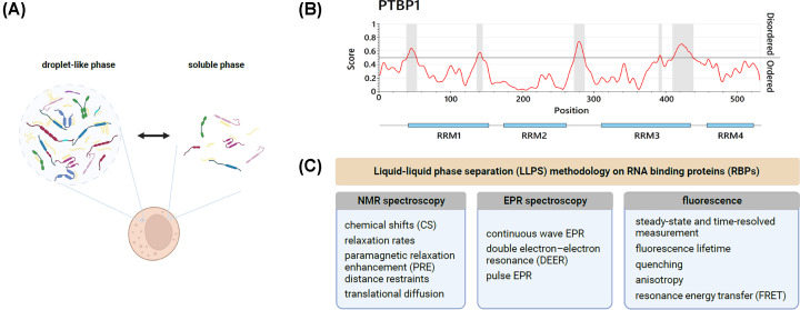
Figure 3. Liquid-liquid phase separation of RNA-binding proteins.
(A) Schematic representation of coexisting, reversible droplet-like (condensed), and soluble (dispersed) phases in the cytoplasm. (B) Prediction of disordered protein regions of the RBP PTBP1. Identified regions (highlighted in grey) are mainly located in flexible linkers, flanking RBDs. Generated using IUPred3 [65]. (C) A summary of commonly exploited methods to study LLPS phenomena involving RBPs in vitro.

