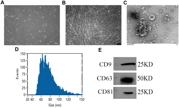
Fig. 1.
Characterization of hAMSC-Exos. A Primary hAMSCs with spindle shapes on culture dishes. B The hAMSCs at passage 3. C The morphology of hAMSC-Exos was captured by TEM. D The diameters and particle concentrations of hAMSC-Exos were detected via NTA. E The specific surface marker proteins (e.g., CD9, CD63, CD81) of hAMSC-Exos were assessed by western blotting

