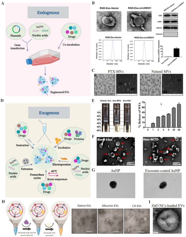
Fig. 2.
Strategies for cargo loading into EVs. A Schemes for the modification of donor cells to release cargos-loaded EVs. B Characterization of exosomes derived from circDIDO1 plasmids-transfected gastric cancer cells.
(Adapted from Guo et al. J Transl Med. 2022;20:326, with permission from Springer Nature [64]) C Transmission electron microscope and scanning electron microscopy for the microvesicles derived from SR4987 cells co-incubated with PTX. (Adapted from Pascucci et al. J Control Release. 2014;192:262–70, with permission from Elsevier [65]) D Schemes for the direct cargo loading into EVs. E Separation of vehicle- and WFA-loaded milk exosomes by Opti-prep density gradient and the release study for Exo-WFA. (Adapted from Munagala et al. Cancer Lett. 2016;371:48–61, with permission from Elsevier [69]) F Transmission electron microscope for drug-loaded exosomes after electroporation. (Adapted from Liang et al. Mol Pharm. 2021;18:1003–13, with permission from American Chemical Society [72]) G Transmission electron microscope for exosome-coated AuNPs after extrusion. (Adapted from Khongkow et al. Sci Rep. 2019;9:8278, with permission from Springer Nature [79]) H Schematic showing the mild sonication procedure and transmission electron microscopy for EVs. (Adapted from Yerneni et al. Acta Biomater. 2022;149:198–212, with permission from Elsevier [75]) I Transmission electron microscopy for ZnO NCs-loaded EVs. (Adapted from Dumontel et al. Cell Biosci. 2022;12:61, with permission from Springer Nature [82])

