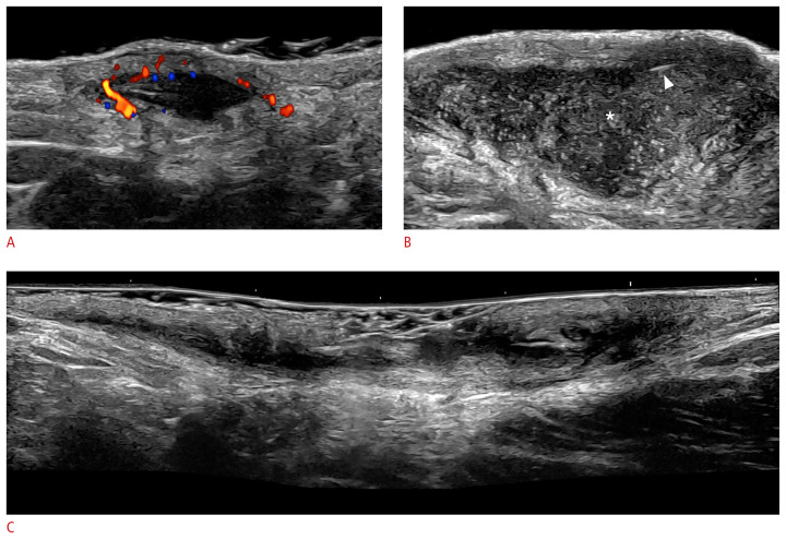
Fig. 13.
Key ultrasound lesions of hidradenitis suppurativa (24 MHz; axillary and groin regions).
Pseudocyst (color Doppler) (A), fluid collection (*) (B), and fistulous tract, also called tunnel (grayscale) (C) are shown. The hypervascularity in the periphery of the pseudocyst in A and the presence of fragments of hair tracts in the fluid collection (B, arrowhead) should be noted. The pseudocyst is in the dermis, and the fluid collection and tunnel are located in the dermis and upper hypodermis.

