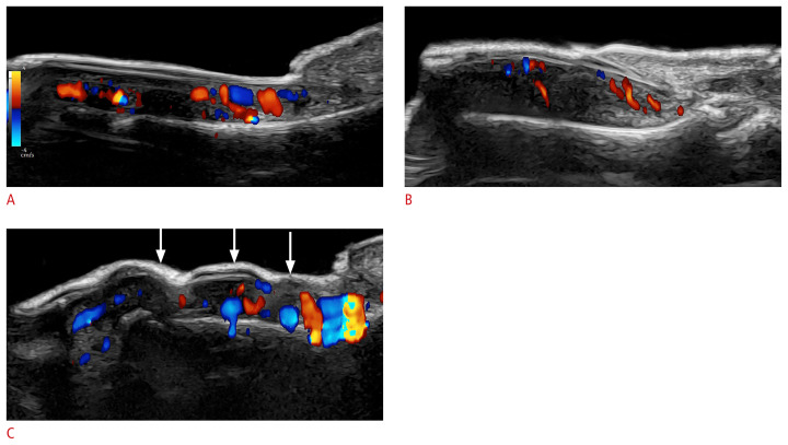
Fig. 20.
Ultrasound patterns of nail psoriasis (color Doppler; 24 MHz; fingernails).
The hypervascularity of the nail beds should be noticed, as well as the morphological alterations. A–C. On ultrasonography, loss of definition of the ventral plate, hyperechoic deposits in the ventral nail plate (A), thickening and decreased echogenicity of the nail bed (B), and a wavy and thick nail plate (C, arrows) are suggestive of nail psoriasis.

