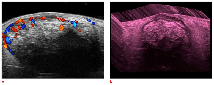
Fig. 5.
Pilomatrixoma of the “target type” (24 MHz; right arm).
Color Doppler (A) and three-dimensional grayscale reconstruction with a color filter (B) show a well-defined, oval-shaped dermal and hypodermal structure. There are multiple hyperechoic focal areas suggestive of calcifications, some of them with posterior acoustic shadowing artifact. On color Doppler, there is hypervascularity in the periphery of the tumor.

