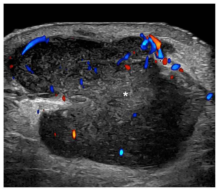
Fig. 9.
Melanoma in-transit metastasis (24 MHz; right arm).
Color Doppler ultrasound demonstrates a hypoechoic hypodermal nodule, slightly heterogeneous with lobulated borders and irregular vascularity. There is increased echogenicity of the surrounding hypodermis. The asterisk marks the metastasis.

