
Figure 7.
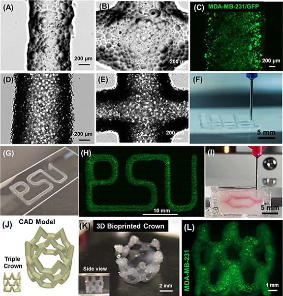
Visual demonstration of bioprinted constructs. Brightfield images showing (A) bioprinted filaments and (B) intersections made of GelMA microgels; (scale: 200 µm). (C) Micrograph of GFP+ MDA-MB-231 cells encapsulated in bioprinted GelMA microgels (scale: 200 µm). Brightfield images of (D) bioprinted filaments, and (E) intersections of GelMA microgels loaded in GelMA. (F) 3D Bioprinting of bilayer grid structures. (G) A photograph and (H) a fluorescent image illustrating cell encapsulating GelMA microgels bioprinted in the shape of ‘PSU,’ the abbreviation of Penn State University. (I) Embedded bioprinting of a branched structure inside a support bath made of Alg microgels. 3D Bioprinted complex-shaped crown constructs with their (J) 3D computer-aided design (CAD) model and (K) macroscopic photographs. (L) GFP+ MDA-MB-231 cells encapsulated in GelMA microgels loaded in bulk GelMA were viable after bioprinting.