Figure 8.

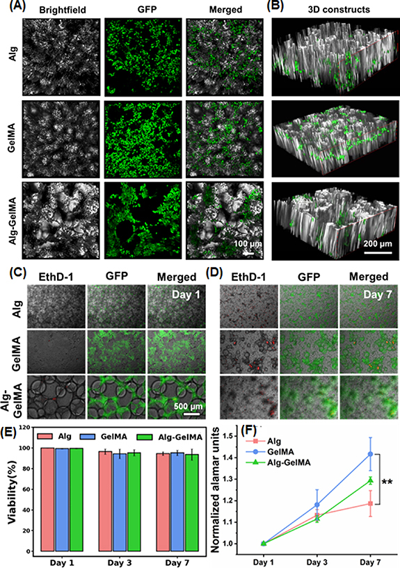
Microscopic images of GFP+ MDA-MB-231 cells seeded on microgels. (A) Images showing GFP+ MDA-MB-231 cells seeded on microgels under brightfield, fluorescence, and merged, and (B) the distribution of these cells in 3D microgel-based constructs. (C) Representative images of GFP+ seeded cells (green) and EthD-1-stained dead cells (red) on Days 1 and 7. (E) Quantification of viability of GFP+ MDA-MB-231 cells seeded on microgels at Days 1, 3, and 7. (D) AlamarBlue assay showing proliferation of GFP+ MDA-MB-231 cells over a week culture (n= 3; p ** < 0.01).
